王兆寅副教授、戴志晖教授课题组在ACS APPLIED MATERIALS & INTERFACES发表研究论文
Formation of a Photoelectrochemical Z-Scheme Structure with Inorganic/Organic Hybrid Materials for Evaluation of Receptor Protein Expression on the Membrane of Cancer Cells
Wang, ZZ (Wang, Zizheng)[ 1,2 ] ; Li, J (Li, Jing)[ 1,2 ] ; Tu, WW (Tu, Wenwen)[ 1,2 ] ; Wang, HS (Wang, Huaisheng)[ 3 ] ; Wang, ZY (Wang, Zhaoyin)[ 1,2 ]*(王兆寅) ; Dai, ZH (Dai, Zhihui)[ 1,2 ]*(戴志晖)
[ 1 ] Nanjing Normal Univ, Sch Chem & Mat Sci, Jiangsu Collaborat Innovat Ctr Biomed Funct Mat, Nanjing 210023, Peoples R China
[ 2 ] Nanjing Normal Univ, Sch Chem & Mat Sci, Jiangsu Key Lab Biofunct Mat, Nanjing 210023, Peoples R China
[ 3 ] Liaocheng Univ, Sch Chem & Chem Engn, Liaocheng 252059, Shandong, Peoples R China
ACS APPLIED MATERIALS & INTERFACES,202006,14(24),26905-26913
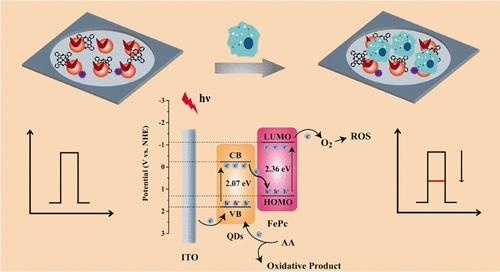
Quantitative analysis of receptor protein expression is essential to give new insights into tumor-related research. Benefitting from their high sensitivity and low background, photoelectrochemical (PEC) platforms are considered as powerful tools for evaluating the expression of receptor proteins. Herein, to reduce the cytotoxicity and facilitate the subsequent assembly, L-cysteine-modified Ag-ZnIn2S4 quantum dots (L-Cys AZIS QDs) are prepared and PEC responses under the irradiation of long wavelength light are obtained. To further improve the PEC behavior, iron phthalocyanine (FePc) is employed to form a Zscheme structure with L-Cys AZIS QDs. The Z-scheme structure based on L-Cys AZIS QDs/FePc hybrid materials exhibits high A photo-to-electric conversion efficiency and can be excited with near-infrared range light. Because hyaluronic acid linked to photoactive materials can recognize CD44 expressed on the membrane of cancer cells, cancer cells are immobilized onto L-Cys AZIS QDs/FePc hybrid materials, inducing a decrease of the photocurrent intensity. Consequently, a PEC cytosensor is constructed to quantify cancer cells expressing CD44. The PEC analytical platform is able to determine A549 cells in the range of 2 X 10(2) to 4.5 X 10(6) cells/mL, and a detection limit of 15 cells/mL is realized in the case of S/N = 3. In addition, the expression of CD44 in A549 and other five cancer cells is measured with this PEC method. Depending on our data, the expression of CD44 in different cancer cells is distinct, indicating great potential of this method in receptor protein-related studies.
文章链接:
https://pubs.acs.org/doi/10.1021/acsami.0c04949
版权与免责声明:本网页的内容由收集互联网上公开发布的信息整理获得。目的在于传递信息及分享,并不意味着赞同其观点或证实其真实性,也不构成其他建议。仅提供交流平台,不为其版权负责。如涉及侵权,请联系我们及时修改或删除。邮箱:sales@allpeptide.com