吴萍副教授和蔡称心教授课题组在TALANTA发表研究论文
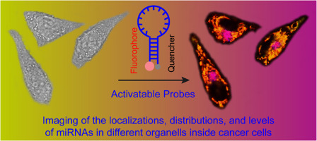
Fluorescence activation imaging of localization, distribution, and level of miRNA in various organelles inside cells
Wu, CL (Wu, Chuanli)[ 1 ] ; Liu, XY (Liu, Xiaoyan)[ 1 ] ; Zheng, YY (Zheng, Yiyi)[ 1 ] ; He, WJ (He, Wenjing)[ 1 ] ; Yang, GC (Yang, Guancao)[ 1 ] ; Wu, P (Wu, Ping)[ 1 ](吴萍)* ; Cai, CX (Cai, Chenxin)[ 1 ](蔡称心)*
[ 1 ] Nanjing Normal Univ, Coll Chem & Mat Sci, Natl & Local Joint Engn Res Ctr Biomed Funct Mat, Jiangsu Collaborat Innovat Ctr Biomed Funct Mat,J, Nanjing 210097, Jiangsu, Peoples R China
TALANTA,201808,186,406-412
This work reports an approach for imaging the localization, distribution, and level of miRNA in different or-ganelles based on an activated fluorescence signal triggered by an alteration of the specific binding-induced conformation of the designed activatable probe. We selected miR-150 as an miRNA example to image its localization, distribution, and level in human cervical cancer cells (HeLa cells). The results indicate that miR-150 is localized and distributed in different subcellular organelles (mainly in mitochondria and lysosomes) and that its levels (actually its concentrations) in lysosomes are higher than those in mitochondria in both HeLa and MCF-7 cells. Moreover, the level of miRNA in cells is displayed in a height-dependent (in z-direction) manner. This approach can also be used to image the localization and distribution of various miRNAs (such as miR-150 and miR-214) in different organelles in cancer cells simultaneously. The probes exhibit high resistance to cellular endo- and exonucleases, with high specificity; the capability of avoiding false signals, with a high signal-to background ratio; and a good ability to operate in complicated environments. The developed approach may provide a useful tool for studying the localization and distribution and evaluating the level of multiple tumor related miRNAs in cells.
版权与免责声明:本网页的内容由收集互联网上公开发布的信息整理获得。目的在于传递信息及分享,并不意味着赞同其观点或证实其真实性,也不构成其他建议。仅提供交流平台,不为其版权负责。如涉及侵权,请联系我们及时修改或删除。邮箱:sales@allpeptide.com