400-998-5282
专注多肽 服务科研
400-998-5282
专注多肽 服务科研

Definition
Herpesvirus has an outer envelope surrounding a capsid and the genomic DNA is a linear double strand, arranged as a toroid wound round a protein plug. Glycoprotein B (gB) is the most conserved glycoprotein of herpesviruses and plays important roles in virus infectivity. Bovine herpesvirus encodes proteins that inhibit specific arms of the immune system. Many synthetic peptides derived from these proteins have shown the ability to inhibit virus replication.
Discovery
Several herpesviruses have been found to induce novel ribonucleotide reductase activities. The herpesviruses enzyme consists of two summits: V136, the large subunit of relative molecular mass (M r) 136,000 (136K) (RR1) and V38, the small subunit (RR2) which forms a complex with the large subunit and is also likely to be essential for enzyme activity. Dutia BM et al., in 1986 identifed a synthetic peptide which specifically inhibits the activity of virus-induced enzyme. They deduced the mechanism of inhibition involves interference with the normal interaction between the two types of subunit 1,2. Okazaki K et al., in 2004 identified amphipathic heptad repeat (HR) regions within gBs of different herpesviruses and demonstrate that a synthetic peptide derived from one of the regions within BoHV-1 gBc bound to the glycoprotein and inhibited the replication of BoHV-1. The peptide did not affect virus entry but interfered with cell-to-cell spread to reduce virus production 3.
Structural Characteristics
Two HR regions were detected in the amino acid sequences of gB of all the herpesviruses analysed. Bovine herpesvirus type 1 (BoHV-1)gB HR1 region commenced 39 aa from the cleavage site and the HR2 region was 22 aa away from the transmembrane (TM) domain. Hydrophobic (I, L, M and V) and aromatic (F, Y and W) amino acids were predominant at the heptad positions of ‘a’ and ‘d’ where the sequences were highly conserved. Helical-wheel representations of the HR1 and HR2 of BoHV-1 gB illustrate the amphipathic property of the regions. In particular, the HR1 region represented a leucine zipper motif 3. Kaposi's Sarcoma-Associated Herpes Virus (KSHV) Protease Substrates, Ac-Val-Tyr-Leu-Lys-Ala-SBzl is used for development of a high throughput colorimetric assay for inhibitor screening 4. HSV-1 Proteinase Substrates,H-His-Thr-Tyr-Leu-Gln-Ala-Ser-Glu-Lys-Phe-Lys-Met-Trp-Gly-NH2 is highly characterized substrate for herpes simplex virus type 1 protease (HSV-1), which is essential for viral nucleocapsid formation and for viral replication 5.
Mode of Action
The ability of bovine herpesvirus type 1 (BHV-1) to impair the immune response can lead to bovine respiratory disease complex (BRDC). Two viral genes, the latency related gene and ORF-E are abundantly expressed during latency, regulate the latency-reactivation cycle. The ability of BHV-1 to enter permissive cells, infect sensory neurons and promote virus spread from sensory neurons to mucosal surfaces following reactivation from latency is also regulated by several viral glycoproteins 6. Infection of cattle with bovine herpesvirus-1 (BHV-1) impairs the cell-mediated immune response (CMI) of the affected host. Interference of BHV-1 with the major histocompatibility complex (MHC) class I antigen presentation pathway was investigated. A considerable down-regulation of the peptide transport activity in bovine epithelial cells, taking place as early as 2 h after virus infection. This down-regulation was also dose-dependent, and, at high multiplicities of infection (moi), led to an almost complete shutdown of TAP. By inhibiting peptide transport into the ER, the virus impairs loading of MHC class I molecules and their subsequent egress from the ER to the cell surface. Thus, BHV-1 selectively interferes with the host's antigen presentation machinery to evade the host's immune response in vivo 7.
Functions
Inhibition of immune system, BHV-1 encodes at least three proteins that can inhibit specific arms of the immune system: (i) bICP0 inhibits interferon-dependent transcription, (ii) the UL41.5 protein inhibits CD8+ T-cell recognition of infected cells by preventing trafficking of viral peptides to the surface of the cells and (iii) glycoprotein G is a chemokine-binding protein that prevents homing of lymphocytes to sights of infection. Following acute infection of calves, BHV-1 can also infect and induce high levels of apoptosis of CD4+ T-cells 6.
Inhibit virus replication, a synthetic peptide derived from the HR region (aa 477–510) of bovine herpesvirus type 1 (BoHV-1) Glycoprotein B (gB) interfered with cell-to-cell spread and consistently inhibited replication of BoHV-1 3.
Antiviral drug, several interactions between virus proteins have been proposed as attractive targets for antiviral drug discovery of herpesvirus, as the exquisite specificity of such cognate interactions affords the possibility of interfering with them in a highly specific and effective manner. Most drugs active against herpesviruses, target the polymerisation activity of the virus DNA polymerase, are limited by pharmacokinetic issues, toxicity and antiviral resistance. A potential novel target for anti-herpesvirus drugs is the interaction between the two subunits of the virus DNA polymerase. 8.
References
Dutia BM, Frame MC, Subak-Sharpe JH, Clark WN, Marsden HS (1986). Specific inhibition of herpesvirus ribonucleotide reductase by synthetic peptides. Nature., 321:439-441.
Frame MC, Marsden HS, Dutia BM (1985). The ribonucleotide reductase induced by herpes simplex virus type 1 involves minimally a complex of two polypeptides (136K and 38K). J Gen Virol., 66:581-587.
Okazaki K, Kida H (2004). A synthetic peptide from a heptad repeat region of herpesvirus glycoprotein B inhibits virus replication. J Gen Virol., 85:2131-2137.
Pray TR, Nomura AM, Pennington MW, Craik CS (1999). Auto-inactivation by cleavage within the dimer interface of Kaposi's sarcoma-associated herpesvirus protease. J. Mol. Biol., 289(2):197-203.
Hall DL, Darke PL (1995). Activation of the herpes simplex virus type 1 protease. J. Biol. Chem., 270(39):22697-22700.
Jonesa C,Chowdhurya S (2007). A review of the biology of bovine herpesvirus type 1 (BHV-1), its role as a cofactor in the bovine respiratory disease complex and development of improved vaccines. Animal Health Research Reviews, 8:187-205.
Hinkley S, Hill AB, Srikumaran S (1998). Bovine herpesvirus-1 infection affects the peptide transport activity in bovine cells. Virus Research., 53(1):91-96.
Loregian A, Palù G (2005). Disruption of the interactions between the subunits of herpesvirus DNA polymerases as a novel antiviral strategy. Clinical Microbiology and Infection, 11(6):437-446.
定义
酶是用于生化反应的非常有效的催化剂。它们通过提供较低活化能的替代反应途径来加快反应速度。酶作用于底物并产生产物。一些物质降低或什至停止酶的催化活性被称为抑制剂。
发现
1965年,Umezawa H分析了微生物产生的酶抑制剂,并分离出了抑制亮肽素和抗痛药的胰蛋白酶和木瓜蛋白酶,乳糜蛋白酶抑制的胰凝乳蛋白酶,胃蛋白酶抑制素抑制胃蛋白酶,泛磷酰胺抑制唾液酸酶,乌藤酮抑制酪氨酸羟化酶,多巴汀抑制多巴胺3-羟硫基嘧啶和多巴胺3-羟色胺酶酪氨酸羟化酶和多巴胺J3-羟化酶。最近,一种替代方法已应用于预测新的抑制剂:合理的药物设计使用酶活性位点的三维结构来预测哪些分子可能是抑制剂1。已经开发了用于识别酶抑制剂的基于计算机的方法,例如分子力学和分子对接。
结构特征
已经确定了许多抑制剂的晶体结构。已经确定了三种与凝血酶复合的高效且选择性的低分子量刚性肽醛醛抑制剂的晶体结构。这三种抑制剂全部在P3位置具有一个新的内酰胺部分,而对胰蛋白酶选择性最高的两种抑制剂在P1位置具有一个与S1特异性位点结合的胍基哌啶基。凝血酶的抑制动力学从慢到快变化,而对于胰蛋白酶,抑制的动力学在所有情况下都快。根据两步机理2中稳定过渡态络合物的缓慢形成来检验动力学。
埃米尔•菲舍尔(Emil Fischer)在1894年提出,酶和底物都具有特定的互补几何形状,彼此恰好契合。这称为“锁和钥匙”模型3。丹尼尔·科什兰(Daniel Koshland)提出了诱导拟合模型,其中底物和酶是相当灵活的结构,当底物与酶4相互作用时,活性位点通过与底物的相互作用不断重塑。
在众多生物活性肽的成熟过程中,需要由其谷氨酰胺(或谷氨酰胺)前体形成N末端焦谷氨酸(pGlu)。游离形式并与底物和三种咪唑衍生抑制剂结合的人QC的结构揭示了类似于两个锌外肽酶的α/β支架,但有多个插入和缺失,特别是在活性位点区域。几种活性位点突变酶的结构分析为针对QC相关疾病5的抑制剂的合理设计提供了结构基础。
作用方式
酶是催化化学反应的蛋白质。酶与底物相互作用并将其转化为产物。抑制剂的结合可以阻止底物进入酶的活性位点和/或阻止酶催化其反应。抑制剂的种类繁多,包括:非特异性,不可逆,可逆-竞争性和非竞争性。可逆抑制剂 以非共价相互作用(例如疏水相互作用,氢键和离子键)与酶结合。非特异性抑制方法包括最终使酶的蛋白质部分变性并因此不可逆的任何物理或化学变化。特定抑制剂 对单一酶发挥作用。大多数毒药通过特异性抑制酶发挥作用。竞争性抑制剂是任何与底物的化学结构和分子几何结构非常相似的化合物。抑制剂可以在活性位点与酶相互作用,但是没有反应发生。非竞争性抑制剂是与酶相互作用但通常不在活性位点相互作用的物质。非竞争性抑制剂的净作用是改变酶的形状,从而改变活性位点,从而使底物不再能与酶相互作用而产生反应。非竞争性抑制剂通常是可逆的。不可逆抑制剂与酶形成牢固的共价键。这些抑制剂可以在活性位点附近或附近起作用。
功能
工业应用中, 酶在商业上被广泛使用,例如在洗涤剂,食品和酿造工业中。蛋白酶用于“生物”洗衣粉中,以加速蛋白质在诸如血液和鸡蛋等污渍中的分解。商业上使用酶的问题包括:它们是水溶性的,这使得它们难以回收,并且一些产物可以抑制酶的活性(反馈抑制)。
药物分子,许多药物分子都是酶抑制剂,药用酶抑制剂通常以其特异性和效力为特征。高度的特异性和效力表明该药物具有较少的副作用和较低的毒性。酶抑制剂在自然界中发现,并且也作为药理学和生物化学的一部分进行设计和生产6。
天然毒物 通常是酶抑制剂,已进化为保护植物或动物免受天敌的侵害。这些天然毒素包括一些已知最剧毒的化合物。
神经气体( 例如二异丙基氟磷酸酯(DFP))通过与丝氨酸的羟基反应生成酯,从而抑制了乙酰胆碱酯酶的活性位点。
参考
1、Scapin G (2006). Structural biology and drug discovery. Curr. Pharm. Des., 12(17):2087–2097.
2、Krishnan R, Zhang E, Hakansson K, Arni RK, Tulinsky A, Lim-Wilby MS, Levy OE, Semple JE, Brunck TK (1998). Highly selective mechanism-based thrombin inhibitors: structures of thrombin and trypsin inhibited with rigid peptidyl aldehydes. Biochemistry, 37 (35):12094-12103.
3、Fischer E (1894). Einfluss der configuration auf die wirkung der enzyme. Ber. Dt. Chem. Ges., 27:2985–2993.
4、Koshland DE (1958). Application of a theory of enzyme specificity to protein synthesis. PNAS., 44 (2):98–104.
5、Huang KF, Liu YL, Cheng WJ, Ko TP, Wang AH (2005). Crystal structures of human glutaminyl cyclase, an enzyme responsible for protein N-terminal pyroglutamate formation. PNAS., 102(37):13117-13122.
6、Holmes CF, Maynes JT, Perreault KR, Dawson JF, James MN (2002). Molecular enzymology underlying regulation of protein phosphatase-1 by natural toxins. Curr Med Chem., 9(22):1981-1989.
Definition
Enzymes are very efficient catalysts for biochemical reactions. They speed up reactions by providing an alternative reaction pathway of lower activation energy. Enzyme acts on substrate and gives rise to a product. Some substances reduce or even stop the catalytic activities of enzymes are called inhibitors.
Discovery
In 1965, Umezawa H analysed enzyme inhibitors produced by microorganisms and isolated leupeptin and antipain inhibiting trypsin and papain, chymostatin inhibiting chymotrypsin, pepstatin inhibiting pepsin, panosialin inhibiting sialidases, oudenone inhibiting tyrosine hydroxylase, dopastin inhibiting dopamine 3-hydroxylase, aquayamycin and chrothiomycin inhibiting tyrosine hydroxylase and dopamine J3-hydroxylase . Recently, an alternative approach has been applied to predict new inhibitors: rational drug design uses the three-dimensional structure of an enzyme's active site to predict which molecules might be inhibitors 1. Computer-based methods for identifying inhibitor for an enzyme have been developed, such as molecular mechanics and molecular docking.
Structural Characteristics
The crystal structures of many inhibitors have been determined. The crystal structures of three highly potent and selective low-molecular weight rigid peptidyl aldehyde inhibitors complexed with thrombin have been determined. All the three inhibitors have a novel lactam moiety at the P3 position, while the two with greatest trypsin selectivity have a guanidinopiperidyl group at the P1 position that binds in the S1 specificity site. The kinetics of inhibition vary from slow to fast with thrombin and are fast in all cases with trypsin. The kinetics are examined in terms of the slow formation of a stable transition-state complex in a two-step mechanism 2.
Emil Fischer in 1894 suggested that both the enzyme and the substrate possess specific complementary geometric shapes that fit exactly into one another.This is known as "the lock and key" model 3. Daniel Koshland suggested induced fit model where substrate and enzymes are rather flexible structures, the active site is continually reshaped by interactions with the substrate as the substrate interacts with the enzyme 4.
N-terminal pyroglutamate (pGlu) formation from its glutaminyl (or glutamyl) precursor is required in the maturation of numerous bioactive peptides. The structure of human QC in free form and bound to a substrate and three imidazole-derived inhibitors reveals an alpha/beta scaffold akin to that of two-zinc exopeptidases but with several insertions and deletions, particularly in the active-site region. The structural analyses of several active-site-mutant enzymes provide a structural basis for the rational design of inhibitors against QC-associated disorders 5.
Mode of Action
Enzymes are proteins that catalyze chemical reactions. Enzymes interact with substrate and convert them into products. Inhibitor binding can stop a substrate from entering the enzyme's active site and/or hinder the enzyme from catalyzing its reaction. There are a variety of types of inhibitors including: nonspecific, irreversible, reversible - competitive and noncompetitive. Reversible inhibitors bind to enzymes with non-covalent interactions like hydrophobic interactions, hydrogen bonds, and ionic bonds. Non-specific methods of inhibition include any physical or chemical changes which ultimately denature the protein portion of the enzyme and are therefore irreversible. Specific Inhibitors exert their effects upon a single enzyme. Most poisons work by specific inhibition of enzymes. A competitive inhibitor is any compound which closely resembles the chemical structure and molecular geometry of the substrate. The inhibitor may interact with the enzyme at the active site, but no reaction takes place. A noncompetitive inhibitor is a substance that interacts with the enzyme, but usually not at the active site. The net effect of a non competitive inhibitor is to change the shape of the enzyme and thus the active site, so that the substrate can no longer interact with the enzyme to give a reaction. Non competitive inhibitors are usually reversible. Irreversible Inhibitors form strong covalent bonds with an enzyme. These inhibitors may act at, near, or remote from the active site .
Functions
Industrial application, enzymes are widely used commercially, for example in the detergent, food and brewing industries. Protease enzymes are used in 'biological' washing powders to speed up the breakdown of proteins in stains like blood and egg. Problems using enzymes commercially include: they are water soluble which makes them hard to recover and some products can inhibit the enzyme activity (feedback inhibition) .
Drug molecules, many drug molecules are enzyme inhibitors and a medicinal enzyme inhibitor is usually characterized by its specificity and its potency. A high specificity and potency suggests that a drug will have fewer side effects and less toxic. Enzyme inhibitors are found in nature and are also designed and produced as part of pharmacology and biochemistry 6.
Natural poisons are often enzyme inhibitors that have evolved to defend a plant or animal against predators. These natural toxins include some of the most poisonous compounds known.
Nerve gases such as diisopropylfluorophosphate (DFP) inhibit the active site of acetylcholine esterase by reacting with the hydroxyl group of serine to make an ester.
References
Scapin G (2006). Structural biology and drug discovery. Curr. Pharm. Des., 12(17):2087–2097.
Krishnan R, Zhang E, Hakansson K, Arni RK, Tulinsky A, Lim-Wilby MS, Levy OE, Semple JE, Brunck TK (1998). Highly selective mechanism-based thrombin inhibitors: structures of thrombin and trypsin inhibited with rigid peptidyl aldehydes. Biochemistry, 37 (35):12094-12103.
Fischer E (1894). Einfluss der configuration auf die wirkung der enzyme. Ber. Dt. Chem. Ges., 27:2985–2993.
Koshland DE (1958). Application of a theory of enzyme specificity to protein synthesis. PNAS., 44 (2):98–104.
Huang KF, Liu YL, Cheng WJ, Ko TP, Wang AH (2005). Crystal structures of human glutaminyl cyclase, an enzyme responsible for protein N-terminal pyroglutamate formation. PNAS., 102(37):13117-13122.
Holmes CF, Maynes JT, Perreault KR, Dawson JF, James MN (2002). Molecular enzymology underlying regulation of protein phosphatase-1 by natural toxins. Curr Med Chem., 9(22):1981-1989.
定义
几种病毒肽是 细胞毒性T淋巴细胞 (CTL)表位或H-2Db限制性表位,有助于 T细胞受体识别过程,并在那里 清除被 病毒感染的细胞。
Discovery
Tsomides TJ等人在1994年建立了一种基于稳定稳定地表达从HIV血清阳性个体获得的HIV-1和CTL的新开发细胞系的检测系统。他们鉴定了MHC-1分子内源性呈递的HIV肽1。可以通过酸洗脱从感染了病毒的细胞中提取此类肽。来自流感病毒核蛋白的天然加工的H–2–Db限制肽和H–2–Kd限制肽均小于相应的合成肽,后者首先用于确定各自的CTL表位。与次要的组织相容性抗原一样,病毒肽的出现似乎严重依赖于MHC I类分子,因为被感染的H-2d细胞不含H-2-Db限制性肽,而被感染的H-2b细胞也不包含HHC-2类。 H-2-Kd限制肽。数据为上述概念提供了直接的实验证据,证明了病毒感染细胞2上与MHC相关的病毒肽的概念。
结构特征
小鼠MHC I类分子(H-2Kb)的X射线结构与两种不同的病毒肽(来自水泡性口炎病毒核蛋白VSV-8和仙台病毒核蛋白SEV-9)复合测定。H-2Kb络合物的VSV-8精制为2.3,SEV-9精制为2.5。H-2Kb的结构与人类HLA I类具有高度相似性,尽管各个结构域的排列可能略有变化。两种肽都以延伸的构象结合,其大部分表面掩埋在H-2Kb结合槽中。纳米单体肽主要通过在其他β构象的中心插入凸起来维持与八聚体相同的氨基和羧基末端相互作用。大多数特异性相互作用是在H-2Kb的侧链原子和肽的主链原子之间。该结合方案在很大程度上解释了以高亲和力结合I类分子的肽序列的巨大多样性。H-2Kb的微小但显着的构象变化与肽结合有关,这些协同运动可能是T细胞受体识别过程不可或缺的一部分3。
行动方式
通过破坏受病毒感染的细胞,受MHC限制的CTL在针对多种病毒的免疫反应中起着核心作用。与抗体不同,CD8 + CTL除了识别来自病毒编码的细胞表面糖蛋白的序列外,还识别来自细胞内病毒蛋白的保守序列。几种病毒表达的基因会下调I类MHC蛋白的表达,从而为它们提供了逃避细胞毒性T淋巴细胞的手段,但使它们易于被NK细胞裂解。I类MHC分子几乎无处不在的表达提供了一种手段,NK细胞可通过这些手段在可能丢失或下调的表面I类MHC分子的修饰靶细胞之间进行区分。I类MHC蛋白的缺失或失调可能部分解释了NK细胞如何调查组织中I类MHC的正常表达,缺失的自我假设。肿瘤细胞下调I类MHC蛋白的表达,而表达I类蛋白的肿瘤细胞则比特异性缺乏I类MHC表达的衍生物更能抵抗NK细胞的杀伤。可以通过转染适当的I类MHC分子来拯救对NK裂解敏感的I类阴性突变细胞系 4,5。
功能
消除病毒感染的细胞,可以通过CTL消除病毒感染的细胞,CTL可以识别与细胞表面2的MHC I类分子结合的病毒衍生肽。
天然杀伤(NK)细胞 被I型主要组织相容性复合物配体的特定同型异型抑制,该配体被多态性抑制受体(例如NKIR1和NKIR2)识别。NK1和NK2特异性克隆识别两组HLA-C同种异型,这些同种异型的特征是在α1螺旋中的残基80(分别为αLys-80和αAsn-80)处具有双态性。谷氨酸脱羧酶或柯萨奇病毒(每种都与自身免疫性糖尿病有关)衍生的肽废除了这种抑制性识别6。
感染后1-2周内会出现针对多种HIV产品(例如gag,pol,nef,env)的CD8 + CTL,这些CTL的异常高频率提示了它们的重要性:通常可以在新鲜分离的中检测到它们没有体外抗原刺激的PBMC通常需要证明其在其他病毒感染中的CTL活性 1。
参考
1、Tsomides TJ, Aldovini A, Johnson RP, Walker BD, Young RA, Eisen HN (1994). Naturally processed viral peptides recognized by cytotoxic T lymphocytes on cells chronically infected by human immunodeficiency virus type 1. J Exp Med., 180(4):1283-1293.
2、Rötzschke O, Falk K, Deres K, Schild H, Norda M, Metzger J, Jung G, Rammensee HG (1990). Isolation and analysis of naturally processed viral peptides as recognized by cytotoxic T cells. Nature, 348(6298):252-254.
3、Fremont DH, Matsumura M, Stura EA, Peterson PA, Wilson IA (1992). Crystal structures of two viral peptides in complex with murine MHC class I H-2Kb. Science, 257(5072):919-927.
4、Ljunggren H-G, Karre K (1990). In search of the 'missing self': MHC molecules and NK cell recognition. Immunol Today., 11(7):237-244.
5、Kärre K, Ljunggren HG, Piontek G, Kiessling R. (1986). Selective rejection of H-2-deficient lymphoma variants suggests alternative immune defence strategy. Nature, 319:675–678.
6、Mandelboim O, Wilson SB, Valés-Gómez M, Reyburn HT, Strominger JL (1997). Self and viral peptides can initiate lysis by autologous natural killer cells. PNAS., 94(9):4604-4609.
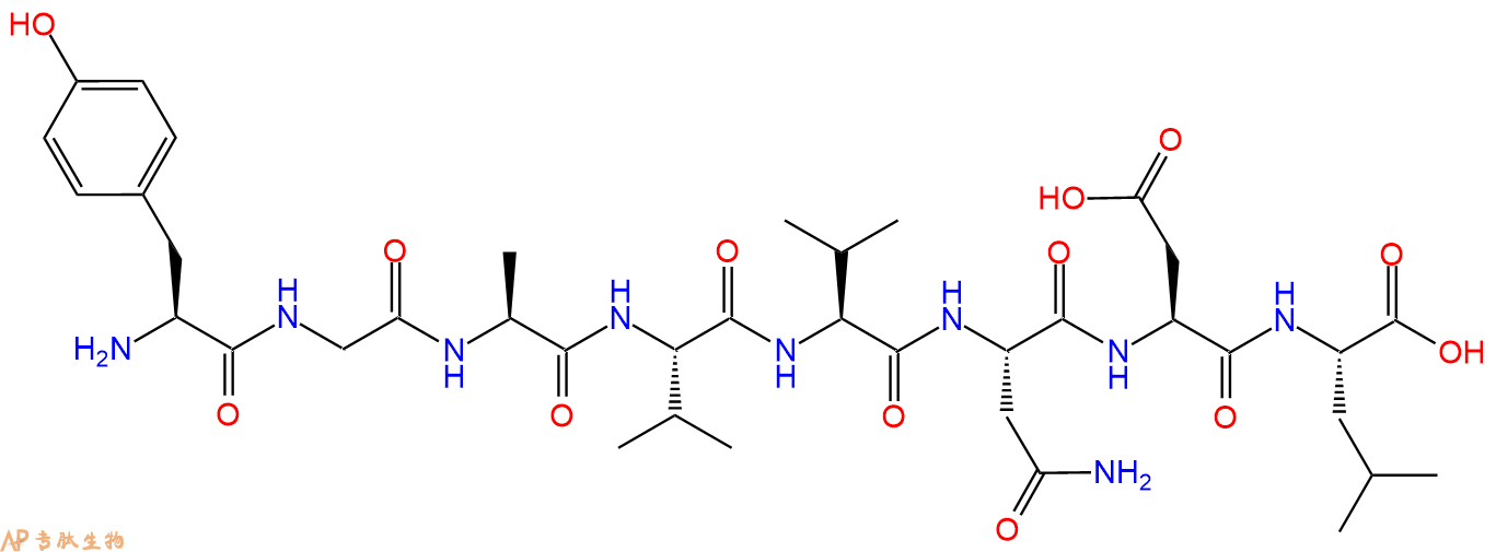
多肽H2N-Tyr-Gly-Ala-Val-Val-Asn-Asp-Leu-COOH的合成步骤:
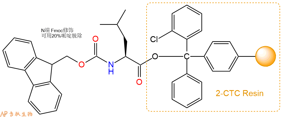
1、合成CTC树脂:称取0.92g CTC Resin(如初始取代度约为0.47mmol/g)和0.52mmol Fmoc-Leu-OH于反应器中,加入适量DCM溶解氨基酸(需要注意,此时CTC树脂体积会增大好几倍,避免DCM溶液过少),再加入1.3mmol DIPEA(Mw:129.1,d:0.740g/ml),反应2-3小时后,可不抽滤溶液,直接加入1ml的HPLC级甲醇,封端半小时。依次用DMF洗涤2次,甲醇洗涤1次,DCM洗涤一次,甲醇洗涤一次,DCM洗涤一次,DMF洗涤2次(这里使用甲醇和DCM交替洗涤,是为了更好地去除其他溶质,有利于后续反应)。得到 Fmoc-Leu-CTC Resin。结构图如下:

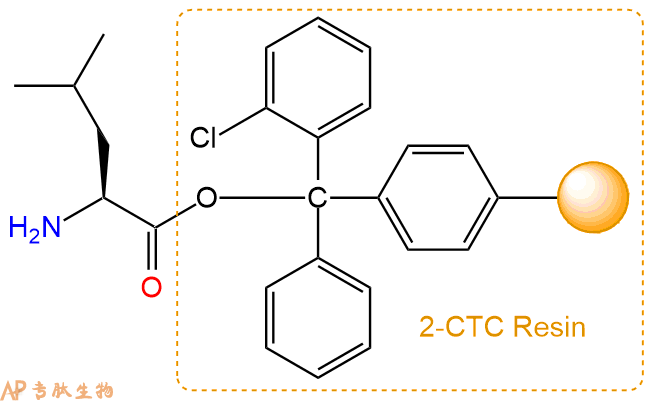
2、脱Fmoc:加3倍树脂体积的20%Pip/DMF溶液,鼓氮气30分钟,然后2倍树脂体积的DMF 洗涤5次。得到 H2N-Leu-CTC Resin 。(此步骤脱除Fmoc基团,茚三酮检测为蓝色,Pip为哌啶)。结构图如下:

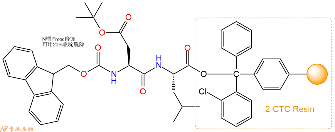
3、缩合:取1.3mmol Fmoc-Asp(OtBu)-OH 氨基酸,加入到上述树脂里,加适当DMF溶解氨基酸,再依次加入2.59mmol DIPEA,1.23mmol HBTU。反应30分钟后,取小样洗涤,茚三酮检测为无色。用2倍树脂体积的DMF 洗涤3次树脂。(洗涤树脂,去掉残留溶剂,为下一步反应做准备)。得到Fmoc-Asp(OtBu)-Leu-CTC Resin。氨基酸:DIPEA:HBTU:树脂=3:6:2.85:1(摩尔比)。结构图如下:

4、依次循环步骤二、步骤三,依次得到
H2N-Asp(OtBu)-Leu-CTC Resin
Fmoc-Asn(Trt)-Asp(OtBu)-Leu-CTC Resin
H2N-Asn(Trt)-Asp(OtBu)-Leu-CTC Resin
Fmoc-Val-Asn(Trt)-Asp(OtBu)-Leu-CTC Resin
H2N-Val-Asn(Trt)-Asp(OtBu)-Leu-CTC Resin
Fmoc-Val-Val-Asn(Trt)-Asp(OtBu)-Leu-CTC Resin
H2N-Val-Val-Asn(Trt)-Asp(OtBu)-Leu-CTC Resin
Fmoc-Ala-Val-Val-Asn(Trt)-Asp(OtBu)-Leu-CTC Resin
H2N-Ala-Val-Val-Asn(Trt)-Asp(OtBu)-Leu-CTC Resin
Fmoc-Gly-Ala-Val-Val-Asn(Trt)-Asp(OtBu)-Leu-CTC Resin
H2N-Gly-Ala-Val-Val-Asn(Trt)-Asp(OtBu)-Leu-CTC Resin
Fmoc-Tyr(tBu)-Gly-Ala-Val-Val-Asn(Trt)-Asp(OtBu)-Leu-CTC Resin
以上中间结构,均可在专肽生物多肽计算器-多肽结构计算器中,一键画出。
最后再经过步骤二得到 H2N-Tyr(tBu)-Gly-Ala-Val-Val-Asn(Trt)-Asp(OtBu)-Leu-CTC Resin,结构如下:

5、切割:6倍树脂体积的切割液(或每1g树脂加8ml左右的切割液),摇床摇晃 2小时,过滤掉树脂,用冰无水乙醚沉淀滤液,并用冰无水乙醚洗涤沉淀物3次,最后将沉淀物放真空干燥釜中,常温干燥24小试,得到粗品H2N-Tyr-Gly-Ala-Val-Val-Asn-Asp-Leu-COOH。结构图见产品结构图。
切割液选择:1)TFA:H2O=95%:5%、TFA:H2O=97.5%:2.5%
2)TFA:H2O:TIS=95%:2.5%:2.5%
3)三氟乙酸:茴香硫醚:1,2-乙二硫醇:苯酚:水=87.5%:5%:2.5%:2.5%:2.5%
(前两种适合没有容易氧化的氨基酸,例如Trp、Cys、Met。第三种适合几乎所有的序列。)
6、纯化冻干:使用液相色谱纯化,收集目标峰液体,进行冻干,获得蓬松的粉末状固体多肽。不过这时要取小样复测下纯度 是否目标纯度。
7、最后总结:
杭州专肽生物技术有限公司(ALLPEPTIDE https://www.allpeptide.com)主营定制多肽合成业务,提供各类长肽,短肽,环肽,提供各类修饰肽,如:荧光标记修饰(CY3、CY5、CY5.5、CY7、FAM、FITC、Rhodamine B、TAMRA等),功能基团修饰肽(叠氮、炔基、DBCO、DOTA、NOTA等),同位素标记肽(N15、C13),订书肽(Stapled Peptide),脂肪酸修饰肽(Pal、Myr、Ste),磷酸化修饰肽(P-Ser、P-Thr、P-Tyr),环肽(酰胺键环肽、一对或者多对二硫键环),生物素标记肽,PEG修饰肽,甲基化修饰肽
以上所有内容,为专肽生物原创内容,请勿发布到其他网站上。


