

400-998-5282
专注多肽 服务科研
400-998-5282
专注多肽 服务科研
MMP-2/MMP-9 Inhibitor III是一种基质金属蛋白酶(MMP-2)和MMP-9的环肽抑制剂
编号:141377
CAS号:244082-19-7
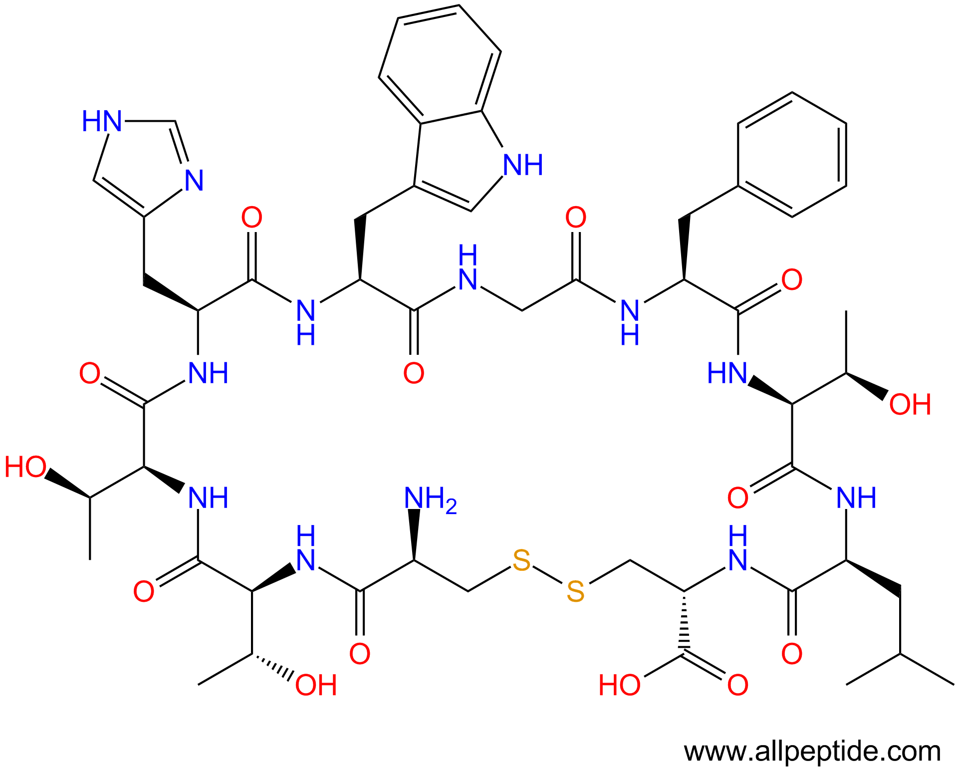
单字母:H2N-CTTHWGFTLC-OH(Disulfide Bridge:C1-C10)
| 编号: | 141377 |
| 中文名称: | 基质金属蛋白酶MMP-2、MMP-9 Inhibitor III、CTT |
| 英文名: | MMP-2/MMP-9 Inhibitor III、CTT |
| CAS号: | 244082-19-7 |
| 单字母: | H2N-CTTHWGFTLC-OH(Disulfide Bridge:C1-C10) |
| 三字母: | H2N-Cys-Thr-Thr-His-Trp-Gly-Phe-Thr-Leu-Cys-OH(Disulfide Bridge:Cys1-Cys10) |
| 氨基酸个数: | 10 |
| 分子式: | C52H71N13O14S2 |
| 平均分子量: | 1166.33 |
| 精确分子量: | 1165.47 |
| 等电点(PI): | - |
| pH=7.0时的净电荷数: | 3.15 |
| 平均亲水性: | -1.2666666666667 |
| 疏水性值: | 0.5 |
| 消光系数: | 5500 |
| 来源: | 人工化学合成,仅限科学研究使用,不得用于人体。 |
| 储存条件: | 负80℃至负20℃ |
| 标签: | 肿瘤(Cancer) 二硫键环肽 抑制剂相关肽(Inhibitor Peptide) 基质金属蛋白酶(MMP) |
| 参考文献(References): | E.Koivunen et al, Nature Biotechnol., 17, 768 (1999) |
MMP-2/MMP-9 Inhibitor III是一种基质金属蛋白酶(MMP-2)和MMP-9的环肽抑制剂。
MMP-2/MMP-9 Inhibitor III is a cyclic peptide inhibitor for matrix metalloproteinases (MMP-2) and MMP-9.
明胶酶抑制剂,但不包括其他MMP家族成员。当与肿瘤靶向性,抗侵袭性相结合时,它成为开发肽模拟抗癌剂的潜在先导化合物。
Inhibitor of gelatinases but not other MMP family members. When combined with tumor targeting, anti-invasive properties, it became a potential lead compound for the development of peptide-mimetic anticancer agents.
CTT肽(CTTHWGFTLC)是一种明胶酶抑制肽,研究发现CTT 肽具有特异性靶向肿瘤的能力。
CTTHWGFTLC靶向MMP-9
化学预防肽是有助于预防疾病(例如癌症或糖尿病)的发作或发展的肽。这些肽可以源自天然来源,例如大豆或牛奶,也可以来自肽模拟物的设计,也可以源自使用合成肽进行的肽筛选。据认为,这些肽中的某些可以充当细胞周期的调节剂,其调节使细胞通过复制周期前进所需的蛋白质的产生和功能。另外,现在有越来越多的证据表明特定的饮食模式,食物和饮料以及其他饮食物质可以而且确实可以预防癌症。越来越多的流行病学研究表明,食物,营养和身体活动在预防和改变癌症过程中很重要。包括植物蛋白酶抑制剂,乳铁蛋白,乳铁蛋白,凝集素和lunasin在内的不同类型的食物蛋白和多肽似乎起着化学预防剂的作用。如今,蛋白质和多肽被认为是一组营养保健品,在预防癌症的不同阶段(包括起始,促进和进展)方面显示出潜力。此外,已经发现在植物中发现的一些蛋白酶抑制剂,例如豆类和大豆,是有效的癌发生抑制剂。致癌作用是引发和促进癌症的过程。 Bowman-Birk抑制剂和Kunitz胰蛋白酶抑制剂就在其中。目前,这些化合物在致癌作用中的生物学功能主要归因于抑制癌细胞的侵袭和转移,但是,其作用机理仍不完全清楚,需要进一步研究以充分阐明它们。
二硫键广泛存在与蛋白结构中,对稳定蛋白结构具有非常重要的意义,二硫键一般是通过序列中的2个Cys的巯基,经氧化形成。
形成二硫键的方法很多:空气氧化法,DMSO氧化法,过氧化氢氧化法等。
二硫键的合成过程, 可以通过Ellman检测以及HPLC检测方法对其反应进程进行监测。
如果多肽中只含有1对Cys,那二硫键的形成是简单的。多肽经固相或液相合成,然后在pH8-9的溶液中进行氧化。
当需要形成2对或2对以上的二硫键时,合成过程则相对复杂。尽管二硫键的形成通常是在合成方案的最后阶段完成,但有时引入预先形成的二硫化物是有利于连合或延长肽链的。通常采用的巯基保护基有trt, Acm, Mmt, tBu, Bzl, Mob, Tmob等多种基团。我们分别列出两种以2-Cl树脂和Rink树脂为载体合成的多肽上多对二硫键形成路线:
二硫键反应条件选择
二硫键即为蛋白质或多肽分子中两个不同位点Cys的巯基(-SH)被氧化形成的S-S共价键。 一条肽链上不同位置的氨基酸之间形成的二硫键,可以将肽链折叠成特定的空间结构。多肽分 子通常分子量较大,空间结构复杂,结构中形成二硫键时要求两个半胱氨酸在空间距离上接近。 此外,多肽结构中还原态的巯基化学性质活泼,容易发生其他的副反应,而且肽链上其他侧链 也可能会发生一系列修饰,因此,肽链进行修饰所选取的氧化剂和氧化条件是反应的关键因素, 反应机理也比较复杂,既可能是自由基反应,也可能是离子反应。
反应条件有多种选择,比如空气氧化,DMSO氧化等温和的氧化过程,也可以采用H2O2,I2, 汞盐等激烈的反应条件。
空气氧化法: 空气氧化法形成二硫键是多肽合成中最经典的方法,通常是将巯基处于还原态的多肽溶于水中,在近中性或弱碱性条件下(PH值6.5-10),反应24小时以上。为了降低分子之间二硫键形成的可能,该方法通常需要在低浓度条件下进行。
碘氧化法:将多肽溶于25%的甲醇水溶液或30%的醋酸水溶液中,逐滴滴加10-15mol/L的碘进行氧化,反应15-40min。当肽链中含有对碘比较敏感的Tyr、Trp、Met和His的残基时,氧化条件要控制的更精确,氧化完后,立即加入维生素C或硫代硫酸钠除去过量的碘。 当序列中有两对或多对二硫键需要成环时,通常有两种情况:
自然随机成环: 序列中的Cys之间随机成环,与一对二硫键成环条件相似;
定点成环: 定点成环即序列中的Cys按照设计要求形成二硫键,反应过程相对复杂。在 固相合成多肽之前,需要提前设计几对二硫键形成的顺序和方法路线,选择不同的侧链 巯基保护基,利用其性质差异,分步氧化形成两对或多对二硫键。 通常采用的巯基保护 基有trt, Acm, Mmt, tBu, Bzl, Mob, Tmob等多种基团。
定义
酶是用于生化反应的非常有效的催化剂。它们通过提供较低活化能的替代反应途径来加快反应速度。酶作用于底物并产生产物。一些物质降低或什至停止酶的催化活性被称为抑制剂。
发现
1965年,Umezawa H分析了微生物产生的酶抑制剂,并分离出了抑制亮肽素和抗痛药的胰蛋白酶和木瓜蛋白酶,乳糜蛋白酶抑制的胰凝乳蛋白酶,胃蛋白酶抑制素抑制胃蛋白酶,泛磷酰胺抑制唾液酸酶,乌藤酮抑制酪氨酸羟化酶,多巴汀抑制多巴胺3-羟硫基嘧啶和多巴胺3-羟色胺酶酪氨酸羟化酶和多巴胺J3-羟化酶。最近,一种替代方法已应用于预测新的抑制剂:合理的药物设计使用酶活性位点的三维结构来预测哪些分子可能是抑制剂1。已经开发了用于识别酶抑制剂的基于计算机的方法,例如分子力学和分子对接。
结构特征
已经确定了许多抑制剂的晶体结构。已经确定了三种与凝血酶复合的高效且选择性的低分子量刚性肽醛醛抑制剂的晶体结构。这三种抑制剂全部在P3位置具有一个新的内酰胺部分,而对胰蛋白酶选择性最高的两种抑制剂在P1位置具有一个与S1特异性位点结合的胍基哌啶基。凝血酶的抑制动力学从慢到快变化,而对于胰蛋白酶,抑制的动力学在所有情况下都快。根据两步机理2中稳定过渡态络合物的缓慢形成来检验动力学。
埃米尔•菲舍尔(Emil Fischer)在1894年提出,酶和底物都具有特定的互补几何形状,彼此恰好契合。这称为“锁和钥匙”模型3。丹尼尔·科什兰(Daniel Koshland)提出了诱导拟合模型,其中底物和酶是相当灵活的结构,当底物与酶4相互作用时,活性位点通过与底物的相互作用不断重塑。
在众多生物活性肽的成熟过程中,需要由其谷氨酰胺(或谷氨酰胺)前体形成N末端焦谷氨酸(pGlu)。游离形式并与底物和三种咪唑衍生抑制剂结合的人QC的结构揭示了类似于两个锌外肽酶的α/β支架,但有多个插入和缺失,特别是在活性位点区域。几种活性位点突变酶的结构分析为针对QC相关疾病5的抑制剂的合理设计提供了结构基础。
作用方式
酶是催化化学反应的蛋白质。酶与底物相互作用并将其转化为产物。抑制剂的结合可以阻止底物进入酶的活性位点和/或阻止酶催化其反应。抑制剂的种类繁多,包括:非特异性,不可逆,可逆-竞争性和非竞争性。可逆抑制剂 以非共价相互作用(例如疏水相互作用,氢键和离子键)与酶结合。非特异性抑制方法包括最终使酶的蛋白质部分变性并因此不可逆的任何物理或化学变化。特定抑制剂 对单一酶发挥作用。大多数毒药通过特异性抑制酶发挥作用。竞争性抑制剂是任何与底物的化学结构和分子几何结构非常相似的化合物。抑制剂可以在活性位点与酶相互作用,但是没有反应发生。非竞争性抑制剂是与酶相互作用但通常不在活性位点相互作用的物质。非竞争性抑制剂的净作用是改变酶的形状,从而改变活性位点,从而使底物不再能与酶相互作用而产生反应。非竞争性抑制剂通常是可逆的。不可逆抑制剂与酶形成牢固的共价键。这些抑制剂可以在活性位点附近或附近起作用。
功能
工业应用中, 酶在商业上被广泛使用,例如在洗涤剂,食品和酿造工业中。蛋白酶用于“生物”洗衣粉中,以加速蛋白质在诸如血液和鸡蛋等污渍中的分解。商业上使用酶的问题包括:它们是水溶性的,这使得它们难以回收,并且一些产物可以抑制酶的活性(反馈抑制)。
药物分子,许多药物分子都是酶抑制剂,药用酶抑制剂通常以其特异性和效力为特征。高度的特异性和效力表明该药物具有较少的副作用和较低的毒性。酶抑制剂在自然界中发现,并且也作为药理学和生物化学的一部分进行设计和生产6。
天然毒物 通常是酶抑制剂,已进化为保护植物或动物免受天敌的侵害。这些天然毒素包括一些已知最剧毒的化合物。
神经气体( 例如二异丙基氟磷酸酯(DFP))通过与丝氨酸的羟基反应生成酯,从而抑制了乙酰胆碱酯酶的活性位点。
参考
1、Scapin G (2006). Structural biology and drug discovery. Curr. Pharm. Des., 12(17):2087–2097.
2、Krishnan R, Zhang E, Hakansson K, Arni RK, Tulinsky A, Lim-Wilby MS, Levy OE, Semple JE, Brunck TK (1998). Highly selective mechanism-based thrombin inhibitors: structures of thrombin and trypsin inhibited with rigid peptidyl aldehydes. Biochemistry, 37 (35):12094-12103.
3、Fischer E (1894). Einfluss der configuration auf die wirkung der enzyme. Ber. Dt. Chem. Ges., 27:2985–2993.
4、Koshland DE (1958). Application of a theory of enzyme specificity to protein synthesis. PNAS., 44 (2):98–104.
5、Huang KF, Liu YL, Cheng WJ, Ko TP, Wang AH (2005). Crystal structures of human glutaminyl cyclase, an enzyme responsible for protein N-terminal pyroglutamate formation. PNAS., 102(37):13117-13122.
6、Holmes CF, Maynes JT, Perreault KR, Dawson JF, James MN (2002). Molecular enzymology underlying regulation of protein phosphatase-1 by natural toxins. Curr Med Chem., 9(22):1981-1989.
Definition
Enzymes are very efficient catalysts for biochemical reactions. They speed up reactions by providing an alternative reaction pathway of lower activation energy. Enzyme acts on substrate and gives rise to a product. Some substances reduce or even stop the catalytic activities of enzymes are called inhibitors.
Discovery
In 1965, Umezawa H analysed enzyme inhibitors produced by microorganisms and isolated leupeptin and antipain inhibiting trypsin and papain, chymostatin inhibiting chymotrypsin, pepstatin inhibiting pepsin, panosialin inhibiting sialidases, oudenone inhibiting tyrosine hydroxylase, dopastin inhibiting dopamine 3-hydroxylase, aquayamycin and chrothiomycin inhibiting tyrosine hydroxylase and dopamine J3-hydroxylase . Recently, an alternative approach has been applied to predict new inhibitors: rational drug design uses the three-dimensional structure of an enzyme's active site to predict which molecules might be inhibitors 1. Computer-based methods for identifying inhibitor for an enzyme have been developed, such as molecular mechanics and molecular docking.
Structural Characteristics
The crystal structures of many inhibitors have been determined. The crystal structures of three highly potent and selective low-molecular weight rigid peptidyl aldehyde inhibitors complexed with thrombin have been determined. All the three inhibitors have a novel lactam moiety at the P3 position, while the two with greatest trypsin selectivity have a guanidinopiperidyl group at the P1 position that binds in the S1 specificity site. The kinetics of inhibition vary from slow to fast with thrombin and are fast in all cases with trypsin. The kinetics are examined in terms of the slow formation of a stable transition-state complex in a two-step mechanism 2.
Emil Fischer in 1894 suggested that both the enzyme and the substrate possess specific complementary geometric shapes that fit exactly into one another.This is known as "the lock and key" model 3. Daniel Koshland suggested induced fit model where substrate and enzymes are rather flexible structures, the active site is continually reshaped by interactions with the substrate as the substrate interacts with the enzyme 4.
N-terminal pyroglutamate (pGlu) formation from its glutaminyl (or glutamyl) precursor is required in the maturation of numerous bioactive peptides. The structure of human QC in free form and bound to a substrate and three imidazole-derived inhibitors reveals an alpha/beta scaffold akin to that of two-zinc exopeptidases but with several insertions and deletions, particularly in the active-site region. The structural analyses of several active-site-mutant enzymes provide a structural basis for the rational design of inhibitors against QC-associated disorders 5.
Mode of Action
Enzymes are proteins that catalyze chemical reactions. Enzymes interact with substrate and convert them into products. Inhibitor binding can stop a substrate from entering the enzyme's active site and/or hinder the enzyme from catalyzing its reaction. There are a variety of types of inhibitors including: nonspecific, irreversible, reversible - competitive and noncompetitive. Reversible inhibitors bind to enzymes with non-covalent interactions like hydrophobic interactions, hydrogen bonds, and ionic bonds. Non-specific methods of inhibition include any physical or chemical changes which ultimately denature the protein portion of the enzyme and are therefore irreversible. Specific Inhibitors exert their effects upon a single enzyme. Most poisons work by specific inhibition of enzymes. A competitive inhibitor is any compound which closely resembles the chemical structure and molecular geometry of the substrate. The inhibitor may interact with the enzyme at the active site, but no reaction takes place. A noncompetitive inhibitor is a substance that interacts with the enzyme, but usually not at the active site. The net effect of a non competitive inhibitor is to change the shape of the enzyme and thus the active site, so that the substrate can no longer interact with the enzyme to give a reaction. Non competitive inhibitors are usually reversible. Irreversible Inhibitors form strong covalent bonds with an enzyme. These inhibitors may act at, near, or remote from the active site .
Functions
Industrial application, enzymes are widely used commercially, for example in the detergent, food and brewing industries. Protease enzymes are used in 'biological' washing powders to speed up the breakdown of proteins in stains like blood and egg. Problems using enzymes commercially include: they are water soluble which makes them hard to recover and some products can inhibit the enzyme activity (feedback inhibition) .
Drug molecules, many drug molecules are enzyme inhibitors and a medicinal enzyme inhibitor is usually characterized by its specificity and its potency. A high specificity and potency suggests that a drug will have fewer side effects and less toxic. Enzyme inhibitors are found in nature and are also designed and produced as part of pharmacology and biochemistry 6.
Natural poisons are often enzyme inhibitors that have evolved to defend a plant or animal against predators. These natural toxins include some of the most poisonous compounds known.
Nerve gases such as diisopropylfluorophosphate (DFP) inhibit the active site of acetylcholine esterase by reacting with the hydroxyl group of serine to make an ester.
References
Scapin G (2006). Structural biology and drug discovery. Curr. Pharm. Des., 12(17):2087–2097.
Krishnan R, Zhang E, Hakansson K, Arni RK, Tulinsky A, Lim-Wilby MS, Levy OE, Semple JE, Brunck TK (1998). Highly selective mechanism-based thrombin inhibitors: structures of thrombin and trypsin inhibited with rigid peptidyl aldehydes. Biochemistry, 37 (35):12094-12103.
Fischer E (1894). Einfluss der configuration auf die wirkung der enzyme. Ber. Dt. Chem. Ges., 27:2985–2993.
Koshland DE (1958). Application of a theory of enzyme specificity to protein synthesis. PNAS., 44 (2):98–104.
Huang KF, Liu YL, Cheng WJ, Ko TP, Wang AH (2005). Crystal structures of human glutaminyl cyclase, an enzyme responsible for protein N-terminal pyroglutamate formation. PNAS., 102(37):13117-13122.
Holmes CF, Maynes JT, Perreault KR, Dawson JF, James MN (2002). Molecular enzymology underlying regulation of protein phosphatase-1 by natural toxins. Curr Med Chem., 9(22):1981-1989.
定义
基质金属蛋白酶(MMP)属于锌内肽酶家族,统称为metzincins。metzincin超家族的特征是高度保守的基序,该基序包含在催化位点与锌结合的三个组氨酸和位于活性位点下方的保守的蛋氨酸。
发现
间质胶原酶,首先被确定的MMP家族成员,最初是在旨在解释designed变态为青蛙1的胶原重塑的实验中发现的。MMP家族由20多个共享相同功能域的相关锌依赖性酶组成。这些酶既具有通常基于优选底物的描述性名称,又具有基于发现顺序的MMP编号系统1。
结构特征
MMP的基本结构由以下同源结构域组成:1)将MMP引导至分泌或质膜插入途径的信号肽;2)前结构域,通过占据活性位点锌使酶具有潜伏期,使底物难以接近催化酶;3)含锌的催化域;4)溶血毒素域,介导与底物的相互作用并赋予酶特异性;5)连接催化和血红蛋白结构域的铰链区。MMP7或基质溶素是最小的MMP,缺少血红素结构域,但在底物降解中表现出特异性。额外的结构域和底物特异性导致MMP分为亚组。
MMP的蛋白质结构中两个序列基序高度保守。在所有MMP的催化域中发现的共有基序HExGHxxGxxH,包含3个与活性中心的锌离子(Zn)配位的组氨酸。PRCGxPD基序位于MMPs前结构域的C末端;该基因座的半胱氨酸残基(C)与活性中心的锌原子的配位赋予了酶2、3潜伏期。
行动方式
MMP的体内活性在几个水平上受到严格控制。这些酶通常以极低的量表达,其转录受到细胞因子和生长因子(如白介素(IL-1,IL-4,IL-6),转化生长因子(EGF,HGF,TGFβ)的正向或负向严格调控。 ),或肿瘤坏死因子α(TNFα的)4, 5。这些调节分子中的一些可以被MMP蛋白水解激活或失活(反馈作用)。转录后,MMP活性受到位于新合成的酶的N末端的前肽所赋予的潜伏期的限制。从细胞分泌后,MMPs的激活取决于前结构域与催化位点的相互作用的破坏,这可能通过前结构域的构象变化或蛋白水解去除而发生。在其前肽中含有弗林蛋白酶样识别结构域的MMP(MMP11,MT-MMP,MMP28)可以通过枯草杆菌蛋白酶丝氨酸蛋白酶家族的成员在反式高尔基体网络中被激活。MMP14在细胞表面的proMMP2激活中起着不可或缺的作用。分泌的MMP的胞外蛋白水解激活可以通过丝氨酸蛋白酶(如纤溶酶)介导,这暗示着这两个酶基团在ECM重塑中具有相互依赖性。某些活动的MMP可以激活其他proMMP,例如MMP9和MMP1的MMP3激活。激活后,MMP会受到内源性抑制剂,自降解和选择性内吞作用的进一步调节。已经证明通过低密度脂蛋白受体相关蛋白(LRP)机制内吞MMP2、9和137。
功能
MMP参与癌症的各个方面-MMP诱导的肝素结合上皮生长因子,胰岛素样生长因子和成纤维细胞生长因子从细胞表面释放(脱落),促进细胞增殖。另一方面,由MMP释放和激活ECM螯合的TGFβ可以导致细胞增殖受到抑制。
炎症性疾病-许多报道暗示MMP1,MMP3和MMP9参与类风湿和骨关节炎。
心血管疾病-大量研究表明,动脉粥样硬化和动脉瘤形成部位6的MMP,尤其是MMP9水平升高。已经提出MMP代表冠状动脉疾病患者中炎症的敏感标志。
肺部疾病- MMP的水平升高已经牵涉在各种肺部疾病,包括急性呼吸窘迫综合征,哮喘,支气管扩张和囊性纤维化的病理生理学。MMP,EMMPRIN和TIMPs是由肺中的许多常驻细胞产生的,因此使它们在疾病中的作用7的分析变得复杂。
中枢神经系统疾病-在观察到MMP9在类似于多发性硬化症和格林-巴利综合症的动物模型中的关键作用之后,MMPs参与了几种不同类型的神经系统疾病。
休克综合征- MMP8和MMP9存储在多形核白细胞的颗粒。这些细胞是炎症和感染过程中的关键效应器。这些MMP在休克中的作用得到了对MMP9缺陷型小鼠的研究的支持,这些小鼠被证明对内毒素休克具有抗性。Dubois等人[ 8]提出,特定的MMP9抑制作用是治疗败血性休克综合症的一种潜在方法。
参考
1. Gross J, Lapiere CM (1962). Collagenolytic activity in amphibian tissues; a tissue culture assay. PNAS., 48:1014-1022.
2. Nagase H, Woessner F (1999). Matrix metalloproteinases. J Biol Chem., 274(31):21491-21494.
3. Birkedal-Hansen H. (1995). Proteolytic remodeling of extracellular matrix. Curr Opin Cell Biol., 7:728-735.
4. Zucker S, Pei D, Cao J, Lopez-Otin C (2003). Membrane type-matrix metalloproteinases (MT-MMP). Cell Surface Proteases., 54:1-74.
5. Yang Z, Strickland DK, Bornstein P (2001). Extracellular MMP-2 levels are regulated by the low-density lipoprotein-related scavenger receptor and thrombospondin. J Biol Chem., 276: 8403-8408.
6. Van den Steen PE, Dubois B, Nelissen I, Rudd PM, Dwek RA, Opdenakker G (2002). Biochemistry and molecular biology of gelatinase B or matrix metalloproteinase-9 (MMP-9). Crit Rev Biochem Mol Biol,. 37:376-536.
7. Haseneen N, Vaday G, Zucker S, Foda HD (2003). Mechanical stretch induces MMP-2 release and activation in lung endothelium: role of EMMPRIN. Am J Physio Lung Cell Mol Physiol., 165:541-547.
8. Dubois B, Starckx S, Pagenstecher A, Oord J, Arnold B, Opdenakker G. Gelatinase B (2002). Deficiency protects against endotoxin shock. Eur J Immunol., 32:2163-2171.
| DOI | 名称 | |
|---|---|---|
| 10.3390/molecules171214230 | Peptide-Based Selective Inhibitors of Matrix Metalloproteinase-Mediated Activities | 下载 |


