

400-998-5282
专注多肽 服务科研
400-998-5282
专注多肽 服务科研
血管紧张素1/2(1-9)TFA包含从血管紧张素I/II肽转化的氨基酸1-9。它是一种抗心肌肥大药物,具有人体代谢产物、大鼠代谢产物、降压药和心脏保护剂的作用。
编号:200053
CAS号:34273-12-6/102816-35-3
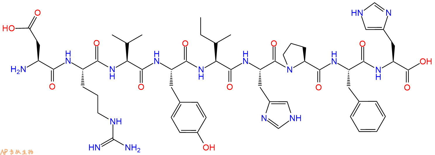
单字母:H2N-DRVYIHPFH-OH
<p>Angiotensin 1/2(1 - 9)是9个氨基酸的肽段(ASP-ARG-VAL-TYR-ILE-HIS-PRO-PHE-HIS)。</p> <p>Angiotensin 1是由肾素作用在肾素血管紧张肽原上形成的,Angiotensin 1具有12个氨基酸,主要由肝脏产生并释放到血液中的α-2-球蛋白。肾素剪切血管紧张肽原上亮氨酸和缬氨酸之间的肽键,产生10个氨基酸肽(Angiotensin 1)。Angiotensin 1主要通过肺内的ACE酶删除两个C端的残基转换为Angiotensin 2(AII)。</p> <p>Angiotensin是一种多肽激素,可以导致血管收缩以及随后的血压增加。Angiotensin还刺激醛固酮的释放,促进远端肾的钠存留引起血压上升。</p> <p>Angiotensin 1/2 (1-9) TFA contains the amino acids 1-9 that are converted from Angiotensin I/II peptide. It is an anti-cardiac hypertrophy drug with the functions of human metabolite, rat metabolite, antihypertensive agent and cardiac protective agent.</p> <p>血管紧张素1/2(1-9)TFA包含从血管紧张素I/II肽转化的氨基酸1-9。它是一种抗心肌肥大药物,具有人体代谢产物、大鼠代谢产物、降压药和心脏保护剂的作用。</p> <p>血管紧张素(1‑9)是中间多肽,用于研究肾素‑血管紧张素级联反应中的连续加工步骤。其结构便于主链取向和电荷分布分析。该片段支持受体结合研究和酶促机制研究。应用包括多肽折叠研究和通路重建。<br /> <br /> Angiotensin (1-9) is an intermediate peptide used to examine sequential processing steps within the renin-angiotensin cascade. Its structure enables analysis of backbone orientation and charge distribution. The fragment supports receptor-binding investigations and enzymatic mechanism research. Applications include peptide folding studies and pathway reconstruction.</p> <p>血管紧张素 I(1-9)三氟乙酸盐
| DOI | 名称 | |
|---|---|---|
| 10.1155/2016/4846819 | Exogenous Angiotensin I Metabolism in Aorta Isolated from Streptozotocin Treated Diabetic Rats | 下载 |
| 10.1016/j.jasms.2005.01.016 | Formation of [b(n-1) + OH + H]+ ion structural analogs by solution-phase chemistry | 下载 |
| 10.1161/01.hyp.18.6.763 | Differential regulation of angiotensin peptide levels in plasma and kidney of the rat | 下载 |
| 10.1002/jms.1544 | Chemical cross-linking with NHS esters: a systematic study on amino acid reactivities | 下载 |
| 10.1177/1470320313498631 | Angiotensin-(1-9) enhances stasis-induced venous thrombosis in the rat because of the impairment of fibrinolysis | 下载 |
| 10.3109/10641969709083179 | Differential regulation of angiotensin peptides in plasma and kidney: effects of adrenalectomy and estrogen treatment | 下载 |
多肽H2N-Asp-Arg-Val-Tyr-Ile-His-Pro-Phe-His-COOH的合成步骤:
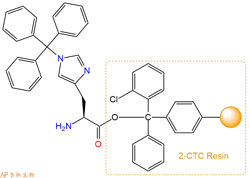
1、合成CTC树脂:称取0.23g CTC Resin(如初始取代度约为0.46mmol/g)和0.13mmol Fmoc-His(Trt)-OH于反应器中,加入适量DCM溶解氨基酸(需要注意,此时CTC树脂体积会增大好几倍,避免DCM溶液过少),再加入0.32mmol DIPEA(Mw:129.1,d:0.740g/ml),反应2-3小时后,可不抽滤溶液,直接加入1ml的HPLC级甲醇,封端半小时。依次用DMF洗涤2次,甲醇洗涤1次,DCM洗涤一次,甲醇洗涤一次,DCM洗涤一次,DMF洗涤2次(这里使用甲醇和DCM交替洗涤,是为了更好地去除其他溶质,有利于后续反应)。得到 Fmoc-His(Trt)-CTC Resin。结构图如下:

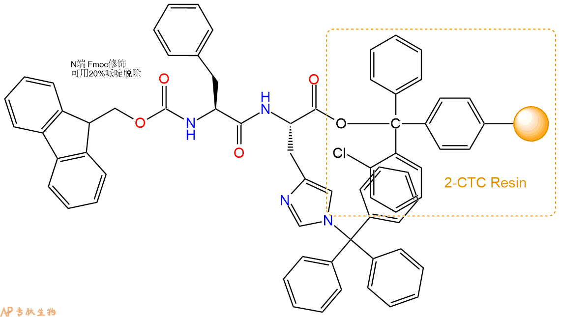
2、脱Fmoc:加3倍树脂体积的20%Pip/DMF溶液,鼓氮气30分钟,然后2倍树脂体积的DMF 洗涤5次。得到 H2N-His(Trt)-CTC Resin 。(此步骤脱除Fmoc基团,茚三酮检测为蓝色,Pip为哌啶)。结构图如下:
3、缩合:取0.32mmol Fmoc-Phe-OH 氨基酸,加入到上述树脂里,加适当DMF溶解氨基酸,再依次加入0.63mmol DIPEA,0.3mmol HBTU。反应30分钟后,取小样洗涤,茚三酮检测为无色。用2倍树脂体积的DMF 洗涤3次树脂。(洗涤树脂,去掉残留溶剂,为下一步反应做准备)。得到Fmoc-Phe-His(Trt)-CTC Resin。氨基酸:DIPEA:HBTU:树脂=3:6:2.85:1(摩尔比)。结构图如下:
4、依次循环步骤二、步骤三,依次得到
H2N-Phe-His(Trt)-CTC Resin
Fmoc-Pro-Phe-His(Trt)-CTC Resin
H2N-Pro-Phe-His(Trt)-CTC Resin
Fmoc-His(Trt)-Pro-Phe-His(Trt)-CTC Resin
H2N-His(Trt)-Pro-Phe-His(Trt)-CTC Resin
Fmoc-Ile-His(Trt)-Pro-Phe-His(Trt)-CTC Resin
H2N-Ile-His(Trt)-Pro-Phe-His(Trt)-CTC Resin
Fmoc-Tyr(tBu)-Ile-His(Trt)-Pro-Phe-His(Trt)-CTC Resin
H2N-Tyr(tBu)-Ile-His(Trt)-Pro-Phe-His(Trt)-CTC Resin
Fmoc-Val-Tyr(tBu)-Ile-His(Trt)-Pro-Phe-His(Trt)-CTC Resin
H2N-Val-Tyr(tBu)-Ile-His(Trt)-Pro-Phe-His(Trt)-CTC Resin
Fmoc-Arg(Pbf)-Val-Tyr(tBu)-Ile-His(Trt)-Pro-Phe-His(Trt)-CTC Resin
H2N-Arg(Pbf)-Val-Tyr(tBu)-Ile-His(Trt)-Pro-Phe-His(Trt)-CTC Resin
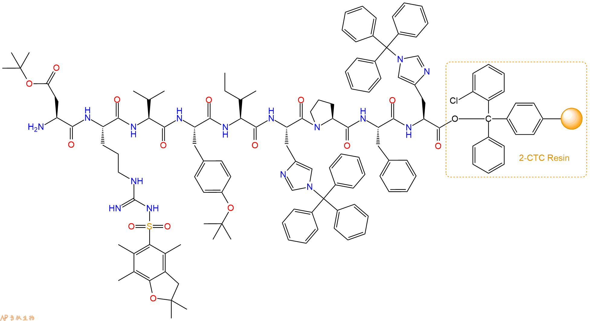
Fmoc-Asp(OtBu)-Arg(Pbf)-Val-Tyr(tBu)-Ile-His(Trt)-Pro-Phe-His(Trt)-CTC Resin
以上中间结构,均可在专肽生物多肽计算器-多肽结构计算器中,一键画出。
最后再经过步骤二得到 H2N-Asp(OtBu)-Arg(Pbf)-Val-Tyr(tBu)-Ile-His(Trt)-Pro-Phe-His(Trt)-CTC Resin,结构如下:
5、切割:6倍树脂体积的切割液(或每1g树脂加8ml左右的切割液),摇床摇晃 2小时,过滤掉树脂,用冰无水乙醚沉淀滤液,并用冰无水乙醚洗涤沉淀物3次,最后将沉淀物放真空干燥釜中,常温干燥24小试,得到粗品H2N-Asp-Arg-Val-Tyr-Ile-His-Pro-Phe-His-COOH。结构图见产品结构图。
切割液选择:1)TFA:H2O=95%:5%、TFA:H2O=97.5%:2.5%
2)TFA:H2O:TIS=95%:2.5%:2.5%
3)三氟乙酸:茴香硫醚:1,2-乙二硫醇:苯酚:水=87.5%:5%:2.5%:2.5%:2.5%
(前两种适合没有容易氧化的氨基酸,例如Trp、Cys、Met。第三种适合几乎所有的序列。)
6、纯化冻干:使用液相色谱纯化,收集目标峰液体,进行冻干,获得蓬松的粉末状固体多肽。不过这时要取小样复测下纯度 是否目标纯度。
7、最后总结:
杭州专肽生物技术有限公司(ALLPEPTIDE https://www.allpeptide.com)主营定制多肽合成业务,提供各类长肽,短肽,环肽,提供各类修饰肽,如:荧光标记修饰(CY3、CY5、CY5.5、CY7、FAM、FITC、Rhodamine B、TAMRA等),功能基团修饰肽(叠氮、炔基、DBCO、DOTA、NOTA等),同位素标记肽(N15、C13),订书肽(Stapled Peptide),脂肪酸修饰肽(Pal、Myr、Ste),磷酸化修饰肽(P-Ser、P-Thr、P-Tyr),环肽(酰胺键环肽、一对或者多对二硫键环),生物素标记肽,PEG修饰肽,甲基化修饰肽
以上所有内容,为专肽生物原创内容,请勿发布到其他网站上。


