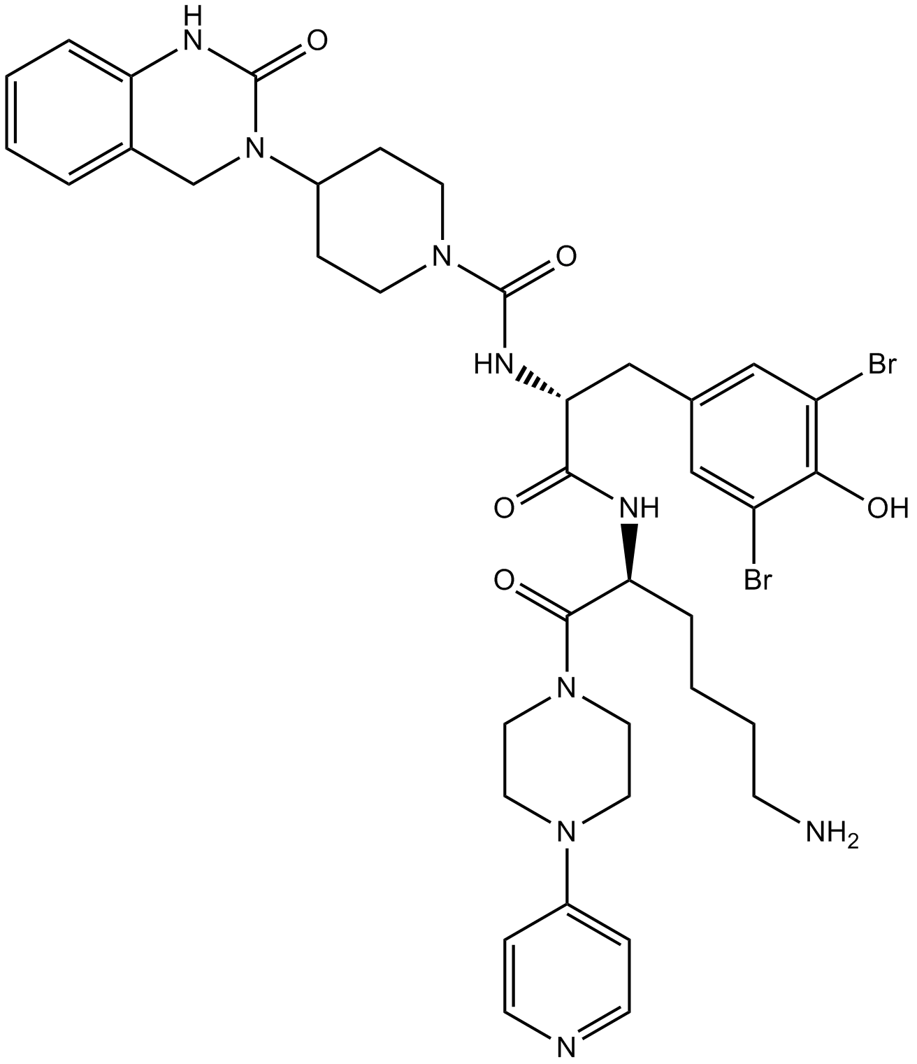
Olcegepant是降钙素基因相关肽1(CGRP 1)的非肽拮抗剂,IC50值为0.03 nM,具有有效性和选择性。
编号:431339
CAS号:204697-65-4
单字母:
| 编号: | 431339 |
| 中文名称: | Olcegepant |
| 英文名: | Olcegepant |
| 英文同义词: | BIBN-4096; BIBN-4096BS;BIBN4096BS; BIBN 4096BS |
| CAS号: | 204697-65-4 |
| 三字母: | N-[(2R)-1-[[(2S)-6-amino-1-oxo-1-(4-pyridin-4-ylpiperazin-1-yl)hexan-2-yl]amino]-3-(3,5-dibromo-4-hydroxyphenyl)-1-oxopropan-2-yl]-4-(2-oxo-1,4-dihydroquinazolin-3-yl)piperidine-1-carboxamide |
| 分子式: | C38H47Br2N9O5 |
| 平均分子量: | 869.66 |
| 标签: | 抑制剂相关肽(Inhibitor Peptide) 降钙素(Calcitonins) |
降钙素基因相关肽(CGRP)的非肽受体,具有有效性和选择性。
IC50:人脑血管为0.1 nM[1]。
Olcegepant是第一个有效的和选择性的非肽类降钙素基因相关肽1(CGRP1)受体拮抗剂,CGRP1是神经性炎性疼痛的关键调节剂。Oehringer Ingelheim GmbH正在研究开发olcegepant,用作治疗急性偏头痛的静脉注射制定治疗剂。
体外实验:SK-N-MC细胞的功能研究表明,CGRP-(8-37)和olcegepant均能抑制CGRP诱导的cAMP产生,pA2值分别为7.8和11.2[1]。
在体实验:用olcegepant预处理(900 mg/kg)抑制了57%的辣椒素(capsaicin)诱导的整个脊三叉神经核的Fos表达。相比之下,在三叉神经节中,olcegepant预处理没有改变磷酸化的胞外信号调节激酶的表达。在中枢神经系统中,CGRP受体抑制可以降低三叉神经脊的活性,而对包括三叉神经节在内的外周神经系统没有影响。这对于未来使用CGRP受体拮抗剂治疗偏头痛可能是重要的[2]。
临床试验:在II期临床试验中,通过对头痛清除率和持续应答率的检测,表明olcegepant减少了60%严重偏头痛患者的头痛。仅观察到了轻度至中度的副作用,且没有不良心血管症状。Olcegepant是一种有效的抗偏头痛药物,耐受性良好,排除使用曲坦和双氢的特定病人,没有血管收缩作用[3]。
定义
酶是用于生化反应的非常有效的催化剂。它们通过提供较低活化能的替代反应途径来加快反应速度。酶作用于底物并产生产物。一些物质降低或什至停止酶的催化活性被称为抑制剂。
发现
1965年,Umezawa H分析了微生物产生的酶抑制剂,并分离出了抑制亮肽素和抗痛药的胰蛋白酶和木瓜蛋白酶,乳糜蛋白酶抑制的胰凝乳蛋白酶,胃蛋白酶抑制素抑制胃蛋白酶,泛磷酰胺抑制唾液酸酶,乌藤酮抑制酪氨酸羟化酶,多巴汀抑制多巴胺3-羟硫基嘧啶和多巴胺3-羟色胺酶酪氨酸羟化酶和多巴胺J3-羟化酶。最近,一种替代方法已应用于预测新的抑制剂:合理的药物设计使用酶活性位点的三维结构来预测哪些分子可能是抑制剂1。已经开发了用于识别酶抑制剂的基于计算机的方法,例如分子力学和分子对接。
结构特征
已经确定了许多抑制剂的晶体结构。已经确定了三种与凝血酶复合的高效且选择性的低分子量刚性肽醛醛抑制剂的晶体结构。这三种抑制剂全部在P3位置具有一个新的内酰胺部分,而对胰蛋白酶选择性最高的两种抑制剂在P1位置具有一个与S1特异性位点结合的胍基哌啶基。凝血酶的抑制动力学从慢到快变化,而对于胰蛋白酶,抑制的动力学在所有情况下都快。根据两步机理2中稳定过渡态络合物的缓慢形成来检验动力学。
埃米尔•菲舍尔(Emil Fischer)在1894年提出,酶和底物都具有特定的互补几何形状,彼此恰好契合。这称为“锁和钥匙”模型3。丹尼尔·科什兰(Daniel Koshland)提出了诱导拟合模型,其中底物和酶是相当灵活的结构,当底物与酶4相互作用时,活性位点通过与底物的相互作用不断重塑。
在众多生物活性肽的成熟过程中,需要由其谷氨酰胺(或谷氨酰胺)前体形成N末端焦谷氨酸(pGlu)。游离形式并与底物和三种咪唑衍生抑制剂结合的人QC的结构揭示了类似于两个锌外肽酶的α/β支架,但有多个插入和缺失,特别是在活性位点区域。几种活性位点突变酶的结构分析为针对QC相关疾病5的抑制剂的合理设计提供了结构基础。
作用方式
酶是催化化学反应的蛋白质。酶与底物相互作用并将其转化为产物。抑制剂的结合可以阻止底物进入酶的活性位点和/或阻止酶催化其反应。抑制剂的种类繁多,包括:非特异性,不可逆,可逆-竞争性和非竞争性。可逆抑制剂 以非共价相互作用(例如疏水相互作用,氢键和离子键)与酶结合。非特异性抑制方法包括最终使酶的蛋白质部分变性并因此不可逆的任何物理或化学变化。特定抑制剂 对单一酶发挥作用。大多数毒药通过特异性抑制酶发挥作用。竞争性抑制剂是任何与底物的化学结构和分子几何结构非常相似的化合物。抑制剂可以在活性位点与酶相互作用,但是没有反应发生。非竞争性抑制剂是与酶相互作用但通常不在活性位点相互作用的物质。非竞争性抑制剂的净作用是改变酶的形状,从而改变活性位点,从而使底物不再能与酶相互作用而产生反应。非竞争性抑制剂通常是可逆的。不可逆抑制剂与酶形成牢固的共价键。这些抑制剂可以在活性位点附近或附近起作用。
功能
工业应用中, 酶在商业上被广泛使用,例如在洗涤剂,食品和酿造工业中。蛋白酶用于“生物”洗衣粉中,以加速蛋白质在诸如血液和鸡蛋等污渍中的分解。商业上使用酶的问题包括:它们是水溶性的,这使得它们难以回收,并且一些产物可以抑制酶的活性(反馈抑制)。
药物分子,许多药物分子都是酶抑制剂,药用酶抑制剂通常以其特异性和效力为特征。高度的特异性和效力表明该药物具有较少的副作用和较低的毒性。酶抑制剂在自然界中发现,并且也作为药理学和生物化学的一部分进行设计和生产6。
天然毒物 通常是酶抑制剂,已进化为保护植物或动物免受天敌的侵害。这些天然毒素包括一些已知最剧毒的化合物。
神经气体( 例如二异丙基氟磷酸酯(DFP))通过与丝氨酸的羟基反应生成酯,从而抑制了乙酰胆碱酯酶的活性位点。
参考
1、Scapin G (2006). Structural biology and drug discovery. Curr. Pharm. Des., 12(17):2087–2097.
2、Krishnan R, Zhang E, Hakansson K, Arni RK, Tulinsky A, Lim-Wilby MS, Levy OE, Semple JE, Brunck TK (1998). Highly selective mechanism-based thrombin inhibitors: structures of thrombin and trypsin inhibited with rigid peptidyl aldehydes. Biochemistry, 37 (35):12094-12103.
3、Fischer E (1894). Einfluss der configuration auf die wirkung der enzyme. Ber. Dt. Chem. Ges., 27:2985–2993.
4、Koshland DE (1958). Application of a theory of enzyme specificity to protein synthesis. PNAS., 44 (2):98–104.
5、Huang KF, Liu YL, Cheng WJ, Ko TP, Wang AH (2005). Crystal structures of human glutaminyl cyclase, an enzyme responsible for protein N-terminal pyroglutamate formation. PNAS., 102(37):13117-13122.
6、Holmes CF, Maynes JT, Perreault KR, Dawson JF, James MN (2002). Molecular enzymology underlying regulation of protein phosphatase-1 by natural toxins. Curr Med Chem., 9(22):1981-1989.
Definition
Enzymes are very efficient catalysts for biochemical reactions. They speed up reactions by providing an alternative reaction pathway of lower activation energy. Enzyme acts on substrate and gives rise to a product. Some substances reduce or even stop the catalytic activities of enzymes are called inhibitors.
Discovery
In 1965, Umezawa H analysed enzyme inhibitors produced by microorganisms and isolated leupeptin and antipain inhibiting trypsin and papain, chymostatin inhibiting chymotrypsin, pepstatin inhibiting pepsin, panosialin inhibiting sialidases, oudenone inhibiting tyrosine hydroxylase, dopastin inhibiting dopamine 3-hydroxylase, aquayamycin and chrothiomycin inhibiting tyrosine hydroxylase and dopamine J3-hydroxylase . Recently, an alternative approach has been applied to predict new inhibitors: rational drug design uses the three-dimensional structure of an enzyme's active site to predict which molecules might be inhibitors 1. Computer-based methods for identifying inhibitor for an enzyme have been developed, such as molecular mechanics and molecular docking.
Structural Characteristics
The crystal structures of many inhibitors have been determined. The crystal structures of three highly potent and selective low-molecular weight rigid peptidyl aldehyde inhibitors complexed with thrombin have been determined. All the three inhibitors have a novel lactam moiety at the P3 position, while the two with greatest trypsin selectivity have a guanidinopiperidyl group at the P1 position that binds in the S1 specificity site. The kinetics of inhibition vary from slow to fast with thrombin and are fast in all cases with trypsin. The kinetics are examined in terms of the slow formation of a stable transition-state complex in a two-step mechanism 2.
Emil Fischer in 1894 suggested that both the enzyme and the substrate possess specific complementary geometric shapes that fit exactly into one another.This is known as "the lock and key" model 3. Daniel Koshland suggested induced fit model where substrate and enzymes are rather flexible structures, the active site is continually reshaped by interactions with the substrate as the substrate interacts with the enzyme 4.
N-terminal pyroglutamate (pGlu) formation from its glutaminyl (or glutamyl) precursor is required in the maturation of numerous bioactive peptides. The structure of human QC in free form and bound to a substrate and three imidazole-derived inhibitors reveals an alpha/beta scaffold akin to that of two-zinc exopeptidases but with several insertions and deletions, particularly in the active-site region. The structural analyses of several active-site-mutant enzymes provide a structural basis for the rational design of inhibitors against QC-associated disorders 5.
Mode of Action
Enzymes are proteins that catalyze chemical reactions. Enzymes interact with substrate and convert them into products. Inhibitor binding can stop a substrate from entering the enzyme's active site and/or hinder the enzyme from catalyzing its reaction. There are a variety of types of inhibitors including: nonspecific, irreversible, reversible - competitive and noncompetitive. Reversible inhibitors bind to enzymes with non-covalent interactions like hydrophobic interactions, hydrogen bonds, and ionic bonds. Non-specific methods of inhibition include any physical or chemical changes which ultimately denature the protein portion of the enzyme and are therefore irreversible. Specific Inhibitors exert their effects upon a single enzyme. Most poisons work by specific inhibition of enzymes. A competitive inhibitor is any compound which closely resembles the chemical structure and molecular geometry of the substrate. The inhibitor may interact with the enzyme at the active site, but no reaction takes place. A noncompetitive inhibitor is a substance that interacts with the enzyme, but usually not at the active site. The net effect of a non competitive inhibitor is to change the shape of the enzyme and thus the active site, so that the substrate can no longer interact with the enzyme to give a reaction. Non competitive inhibitors are usually reversible. Irreversible Inhibitors form strong covalent bonds with an enzyme. These inhibitors may act at, near, or remote from the active site .
Functions
Industrial application, enzymes are widely used commercially, for example in the detergent, food and brewing industries. Protease enzymes are used in 'biological' washing powders to speed up the breakdown of proteins in stains like blood and egg. Problems using enzymes commercially include: they are water soluble which makes them hard to recover and some products can inhibit the enzyme activity (feedback inhibition) .
Drug molecules, many drug molecules are enzyme inhibitors and a medicinal enzyme inhibitor is usually characterized by its specificity and its potency. A high specificity and potency suggests that a drug will have fewer side effects and less toxic. Enzyme inhibitors are found in nature and are also designed and produced as part of pharmacology and biochemistry 6.
Natural poisons are often enzyme inhibitors that have evolved to defend a plant or animal against predators. These natural toxins include some of the most poisonous compounds known.
Nerve gases such as diisopropylfluorophosphate (DFP) inhibit the active site of acetylcholine esterase by reacting with the hydroxyl group of serine to make an ester.
References
Scapin G (2006). Structural biology and drug discovery. Curr. Pharm. Des., 12(17):2087–2097.
Krishnan R, Zhang E, Hakansson K, Arni RK, Tulinsky A, Lim-Wilby MS, Levy OE, Semple JE, Brunck TK (1998). Highly selective mechanism-based thrombin inhibitors: structures of thrombin and trypsin inhibited with rigid peptidyl aldehydes. Biochemistry, 37 (35):12094-12103.
Fischer E (1894). Einfluss der configuration auf die wirkung der enzyme. Ber. Dt. Chem. Ges., 27:2985–2993.
Koshland DE (1958). Application of a theory of enzyme specificity to protein synthesis. PNAS., 44 (2):98–104.
Huang KF, Liu YL, Cheng WJ, Ko TP, Wang AH (2005). Crystal structures of human glutaminyl cyclase, an enzyme responsible for protein N-terminal pyroglutamate formation. PNAS., 102(37):13117-13122.
Holmes CF, Maynes JT, Perreault KR, Dawson JF, James MN (2002). Molecular enzymology underlying regulation of protein phosphatase-1 by natural toxins. Curr Med Chem., 9(22):1981-1989.
Definition
Calcitonin is a 32-amino acid linear polypeptide hormone produced primarily by the parafollicular cells in humans and ultimobranchial body in many other animals1. It acts to reduce blood calcium (Ca2+), opposing the effects of parathyroid hormone (PTH). Calcitonin is a product of the CALC1 gene and is initially produced as a precursor1.
Discovery
Calcitonin was purified in 1962 by Copp and Cheney2. While it was initially considered a secretion of the parathyroid glands, it was later identified as the secretion of the C-cells of the thyroid gland 3 .
Classification
CALC1 gene belongs to a superfamily of related protein hormone precursors that includes islet amyloid precursor protein, calcitonin gene-related peptide, and the precursor of adrenomedullin 4 .
Structural Characteristics
Human calcitonin is a 32 amino acid peptide and is formed from procalcitonin (Cleavage products: Calcitonin, Katalin and a protein fragment)5. It has an N-terminal disulphide bridge and a C-terminal proline amide residue, shown to potently inhibit bone resorption5. Alternative splicing of the gene coding for calcitonin produces a distantly related peptide of 37 amino acids, called calcitonin gene-related peptide (CGRP) 5.
Mode of action
Calcitonin exerts its functions by binding to calcitonin receptor that is a G-protein coupled receptor. Upon binding, the receptor triggers the formation of cAMP, a second messenger which in turn activates various signaling pathways in the target cell (Eg: Osteoblasts) 6 .
Functions
Calcitonin is mainly involved in the metabolism of Ca and phosphorous in the cell. Calcitonin secretion is stimulated by rise in Ca levels in the body. It inhibits Ca intake by the intestine and also prevent loss of Ca from the bones during pregnancy and lactation7It also inhibits osteoclast activity in the bones8. This property of calcitonin is utilized for treatment of osteoporosis and osteoarthritis and recently has been tried for bone metastasis1.Procalcitonin is released during severe infection where it is involved in Ca homeostasis. It is also used as a marker for sepsis8.
References
1. Inzerillo AM, Zaidi M, Huang CL (2004). Calcitonin: physiological actions and clinical applications. J Pediatr Endocrinol Metab., 17(7), 931-40.
2. Copp DH, Cheney B (1962). Calcitonin-a hormone from the parathyroid which lowers the calcium-level of the blood. Nature, 193, 381–2.
3. Stevenson JC, Evans IM (1981). Pharmacology and therapeutic use of calcitonin. Drugs, 21(4), 257-72.
4. Zaidi M, Inzerillo AM, Moonga BS, Bevis PJ, Huang CL (2002). Forty years of calcitonin--where are we now? A tribute to the work of Iain Macintyre, FRS, Bone, 30(5), 655-63.
5. Andreotti G, Méndez BL, Amodeo P, Morelli MA, Nakamuta H, Motta A (2006). "Structural determinants of salmon calcitonin bioactivity: the role of the Leu-based amphipathic alpha-helix". J. Biol. Chem., 281 (34), 24193–203.
6. Purdue BW, Tilakaratne N, Sexton PM (2002). Molecular pharmacology of the calcitonin receptor. Recept. Channels, 8 (3-4), 243–55.
7. Woodrow JP, Sharpe CJ, Fudge NJ, Hoff AO, Gagel RF, Kovacs CS (2006). Calcitonin plays a critical role in regulating skeletal mineral metabolism during lactation. Endocrinology, 147(9), 4010-21.
8. BalcI C, Sungurtekin H, Gürses E, Sungurtekin U, Kaptanoglu B (2003). Usefulness of procalcitonin for diagnosis of sepsis in the intensive care unit. Crit Care, 7 (1), 85–90
Definition
Calcitonin gene-related peptide (CGRP) is a 37-amino acid neuropeptide with potent receptor mediated vasodilatory and cardioexcitatory properties1.
Discovery
It was discovered when alternative processing of RNA transcripts from the calcitonin gene were shown to result in the production of distinct mRNAs encoding CGRP2. A human form of CGRP was isolated from thyroid tissue of patients with medullary thyroid carcinoma3.
Classification
CGRP belongs to the regulatory-peptide family that also includes adrenomedullin and amylin4.
Structural Characteristics
CGRP consists of an amino-terminal disulphide bridge linked loop between amino acids 2 and 7 followed by alpha helix between amino acids 8 and 18 and a poorly defined turn between residues 19 and 215. The carboxy and amino terminals of CGRP can interact independently with its receptors5.
Mode of action
CGRP exerts its function by binding to two G-protein coupled receptors, CGRP1 and CGRP2. One of the major functions of CGRP is vasodilation of cardiac muscles5. In order to achieve this, CGRP first binds to CGRP1 receptor which results in the production of cAMP which in turn activates Protein Kinase A (PKA)6. PKA phosphorylates and opens potassium channels that cause relaxation of muscles6.
Functions
CGRP is widely distributed in the central and peripheral nervous systems5. It produces vascular relaxation via binding to CGRP1 receptor5. Studies in mice have shown that CGRP may play a role in controlling blood pressure5. CGRP also protects tissue injury through its vasodilatory functions. Through its activity as a vasodilator, CGRP influence the activity of inflammatory cells by recruiting more cells at the site of inflammation7. CGRP plays a role in migraine as it is found that its levels raise during painful phases of the disease8. CGRP plays a protective role in cardiac tissue. The infusion of CGRP is beneficial in increasing cardiac output and lowering blood pressure in patients with congestive heart failure5.
References
1. Tortorella C, Macchi C, Forneris M and Nussdorfer GG (2001). Calcitonin gene-related peptide (CGRP), acting via CGRP type 1 receptors, inhibits potassium-stimulated aldosterone secretion and enhances basal catecholamine secretion from rat adrenal gland. Int. J Mol. Med., 8(3), 261-4.
2. Amara SG, Jonas V, Rosenfeld MG, Ong ES and Evans RM (1982). Alternative RNA processing in calcitonin gene expression generates mRNAs encoding different polypeptide products. Nature, 298, 240–244.
3. Aiyar N, Rand K, Elshourbagy NA, Zeng Z, Adamou JE, Bergsma DJ, and Li Y (1996). A cDNA encoding the Calcitonin Generelated peptide type 1 receptor. J Biol Chem., 271, 11325–11329.
4. Bell D and McDermott BJ (1996). Calcitonin gene-related peptide in the cardiovascular system: characterization of receptor populations and their (patho)physiological significance. Pharmacol Rev., 48, 253–288.
5. Susan DB and Andrew DG (2004). Vascular Actions of Calcitonin Gene-Related Peptide and Adrenomedullin. Physiol Rev., 84, 903-934.
6. Hirata Y, Takagi Y, Takata S, Fukuda Y, Yoshimi H, and Fujita T (1988). Calcitonin gene-related peptide receptor in cultured vascular smooth muscle and endothelial cells. Biochem Biophys Res Commun., 151, 1113–1121.
7. Lambrecht BN (2001). Immunologists getting nervous: neuropeptides, dendritic cells and T cell activation. Respir Res., 2, 133–138.
8. Durham, P (2006). Calcitonin gene-related peptide (CGRP) and migraine. Headache, 48: S3–8.


